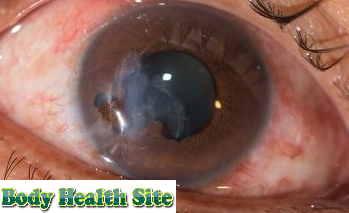
Hifema is the accumulation of blood in the front chamber of the eye between the cornea (the clear membrane of the eye) and the iris (the rainbow membrane). Hifema usually occurs due to injury or trauma that causes the iris or pupil to tears. This bleeding is different from bleeding which occurs in a thin layer above the white part of the eye (conjunctiva), which is known as a subconjunctival hemorrhage. A subconjunctival hemorrhage is not dangerous and does not cause pain, whereas hifema usually feels very painful.
This collection of blood in hifema can cover half of the vision or obstruct overall vision. Conditions that can be experienced by adults and children need to get medical help as soon as possible to prevent permanent damage to vision.
Symptoms of Hifema
A common symptom of hyphae that is seen is bleeding in the front chamber of the eye. If bleeding is still small, hifema cannot be seen in plain view. But if there is a lot of bleeding, the eyes can look like they are filled with blood.
 |
| Hifema, Definition, Symptoms, Causes, Diagnosis, Treatment, Complications |
In addition, hyphae can also cause symptoms such as:
- Eye pain.
- Blurred or obstructed vision.
- Sensitive to light.
Based on the amount of blood, hifema can be divided into 4 levels. The first level is indicated by bleeding less than one-third of the front chamber. The second level occurs when blood fills one third to one half of the front of the eye. At the third level, the blood layer meets half of the front chamber of the eye. Whereas the fourth level is characterized by bleeding that fills the entire anterior chamber, which looks like a number 8 billiard ball or also called a ball hifema 8. However, most of the hyphema that occurs is at the first level. Apart from these four levels, there is also bleeding in the anterior chamber which is not visible to the naked eye. This condition is called microhifema.
Causes of Hifema
Some conditions that can be the cause of the occurrence of hifema, including:
- Injury to the eye, due to an accident or during exercise, for example, exposed to a badminton shuttlecock to the eye.
- Blood vessels on the surface of the iris are not normal.
- Eye infections due to the herpes virus.
- Blood clotting disorders, such as hemophilia and sickle cell anemia.
- Postoperative eye complications.
- Spontaneous hifema can occur due to vascular abnormalities or new blood vessel formation (neovascularization), eye cancer, and uveitis.
Hifema diagnosis
To diagnose hyphema, the ophthalmologist will ask the patient about a history of eye injuries and a physical examination of the eye. Eye examination involves examining the condition of the inside of the eye using a tool called a slit lamp. The doctor will also check the pressure on the eyeball and the sharpness of vision. If a severe injury occurs, the doctor can request a CT scan of the eye.
Hifema treatment
Treatment of hifema is determined by considering the age and overall health condition. Mild hifema can heal on its own within 1 week. Nevertheless. home care must still be carried out based on the eye doctor's instructions because some people with hifema can experience bleeding again if the treatment is not carried out properly.
Things that will be advised by the doctor to prevent the hifema from getting worse, including:
- Reducing severe physical activity and taking a lot of rest.
- Limit eye movements.
- Wear special eye protection to prevent injury.
- Supporting the head when sleeping, at least with a slope of 40 degrees, so that blood can be absorbed into the body.
In addition, doctors will recommend taking painkillers, such as paracetamol, if needed. For aspirin use and nonsteroidal anti-inflammatory drugs (naproxen or ibuprofen), it is not recommended because it can trigger further bleeding.
In addition, doctors can also give eye drops containing corticosteroids to reduce inflammation and drops to dilate the pupils to relieve pain. Vomiting prevention drugs can sometimes be given to patients with hifema because vomiting can increase pressure on the eyes.
During continued care at home, patients must always monitor the condition of pressure on the eyes. Pressure on the eyeball can increase due to red blood cells that block the flow of fluid in the eyeball. If the pressure on the eye increases or bleeding returns, the patient must be hospitalized. The same conditions apply to children and the elderly who cannot follow care instructions independently at home. Hospital care is needed to monitor the condition of hifema more carefully.
Whereas for micro-hyphema sufferers, an examination with an ophthalmologist needs to be done every 5 days to observe the sharpness of vision, eye pressure, and the condition of the front chamber.
Complications of Hifema
Hyphema sufferers are at risk of experiencing increased eye pressure which causes glaucoma. This condition is a chronic disease that can lead to blindness.
Related Search:
- hifema,
- hifema ocular,
- hifema tratamiento,
- hifema definicion,
- icd 10 hifema,
- hifema grading,
- hifema e hiposfagma,
- hifema in eye,


